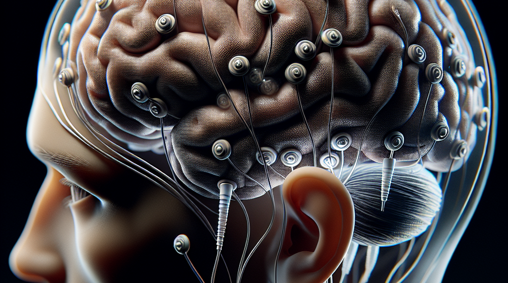
When someone suffers a stroke, the road to recovery can be long and uncertain. A stroke disrupts the brain’s blood supply, which can lead to the death of brain cells and a range of motor deficits, such as partial paralysis or weakness on one side of the body. However, the brain has a remarkable ability to reorganize itself—a process known as neuroplasticity. This ability is central to recovery, and understanding it better could lead to improved treatments. A new study has shed light on this process by using a combination of Transcranial Magnetic Stimulation (TMS) and Electroencephalography (EEG) to track changes in the brain’s activity and connections in stroke patients.
TMS-EEG: A Window into the Brain’s Recovery
TMS is a technique that uses magnetic fields to stimulate small regions of the brain, while EEG records the brain’s electrical activity through electrodes placed on the scalp. By combining these two methods, researchers can observe how different areas of the brain respond and communicate with each other after a stroke. In this study, TMS-EEG was used to measure changes in signal processing and network alterations in 40 stroke patients, from the acute phase immediately following the stroke to the chronic phase, which can last for months or years.
The Significance of Slow-Wave Responses
The researchers discovered that slow-wave responses to TMS, similar to those observed during deep sleep, were present in patients with severe motor deficits early after their stroke. These slow waves are a sign of cortical bistability, a state where the brain alternates between active and silent phases. This condition was linked to reduced complexity in the brain’s signals and was a predictor of less favorable motor outcomes. Interestingly, these slow-wave responses were not permanent; they disappeared in the chronic phase of recovery, suggesting that they could be an early indicator of poor recovery but not a fixed state.
Predicting Recovery and Guiding Treatment
One of the most promising aspects of this research is the potential for TMS-EEG to predict motor recovery. By analyzing the initial signals and the degree of motor deficit, clinicians might be able to forecast a patient’s recovery trajectory. This could be incredibly valuable for tailoring rehabilitation strategies to individual needs. Moreover, the study found that lesions affecting brainstem fibers and leading to disconnection of the pedunculopontine tegmental nucleus were associated with the slow-wave responses. This finding highlights the role of structural disconnection in impaired recovery and suggests that targeting these areas could be a new avenue for treatment.
Looking to the Future
The implications of this study are far-reaching. TMS-EEG could become a crucial tool in monitoring the reorganization of the motor system over time, guiding interventions, and improving functional recovery after a stroke. As we continue to unravel the complexities of the brain’s response to injury, these insights offer hope for developing more effective rehabilitation methods that could significantly enhance the quality of life for stroke survivors. With continued research, the future for stroke recovery looks increasingly optimistic, with the potential for personalized treatments that are informed by the brain’s own signals of healing and adaptation.


